TTA Surgery
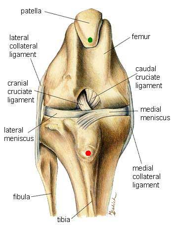
The Normal KneeThe normal Knee Joint (also known as the Stifle joint), has multiple structures that are important to its function. This drawing shows a view from the front with the muscles removed. It is important to note that the Patellar Tendon, a vital structure in the joint has been removed so that you can see "behind" it. The Patellar Tendon is a thick, tough band that runs from the Patella (green dot) to the Tibial Tuberosity (red dot). |
 |
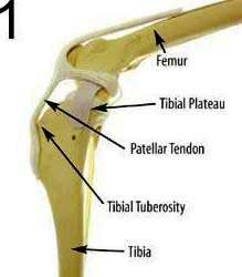
Normal JointThe normal joint, viewed from the side, shows the upper bone, the femur, and the lower bone, the tibia. The Tibial Plateau is the actual point of contact between the femur and the tibia. In this diagram the Patellar Tendon is visible. It is this structure that must offset the abnormal forces that are created by a rupture of the cranial cruciate ligament. |
 |
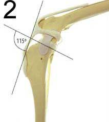
Typical Joint AngleIn the typical joint, the angle formed between the Tibial Plateau and the Patellar Tendon is about 115 degrees when the leg is in a normal standing position. |
 |
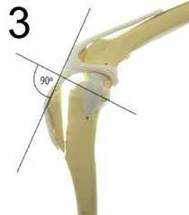
Corrected AngleThe abnormal motion that occurs in a knee with a torn cruciate ligament is called Tibial Thrust. After the TTA Surgery, the corrected angle is now 90 degrees, which will offset the forces in the knee that tend to make it unstable. |
 |
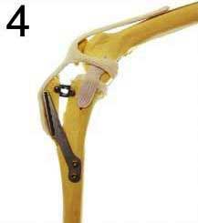
Surgical AppearanceThis diagram shows the knee once it has been stabilized with the appropriate Titanium implants. These implants are very lightweight and are designed to stay in permanently. |
 |